Doravirine placental cotyledon perfusion (Bukkems 2022)
Source:vignettes/articles/Bukkems_2022_doravirine_placentaperfusion.Rmd
Bukkems_2022_doravirine_placentaperfusion.RmdModel and source
#> ℹ parameter labels from comments will be replaced by 'label()'Citation: Bukkems VE, van Hove H, Roelofsen D, Freriksen JJM, van Ewijk-Beneken Kolmer EWJ, Burger DM, van Drongelen J, Svensson EM, Greupink R, Colbers A. Prediction of Maternal and Fetal Doravirine Exposure by Integrating Physiologically Based Pharmacokinetic Modeling and Human Placenta Perfusion Experiments. Clin Pharmacokinet. 2022;61:1129-1141. doi:10.1007/s40262-022-01127-0. PMCID: PMC9349081. Structure: Section 2.2, Figure 1A and Equations 1, 2, 4, 6 and 7; fixed inputs: Table 1; final estimates: Table 2; cotyledon weights: Electronic Supplementary Material Online Resource 4.
Open-access full text: https://europepmc.org/article/MED/PMC9349081
What these files are, and what they are not
Bukkems and colleagues built two kinds of model:
| # | Model | Implementation | In this package? |
|---|---|---|---|
| 1 | Ex vivo cotyledon perfusion model (Section 2.2, Figure 1A, Equations 1-7, Tables 1-2; closed-open re-fit in ESM Online Resources 13-19) | NONMEM 7.4 | yes |
| 2 | Whole-body maternal-fetal pregnancy PBPK model with a permeability-limited placenta (ESM Online Resource 10) | Simcyp V20 | no |
Model 2 is a Simcyp platform model: its organ volumes and blood
flows, tissue partition coefficients and CYP3A4 reference concentrations
are computed inside the Simcyp pregnancy population database and are not
printed in the paper, so it cannot be reconstructed from the
publication. What transfers between the two is a pair of numbers: the
fitted maternal- and fetal-facing transfer clearances,
CLpdm and CLpdf, which the authors converted
to 0.0507 and 0.0075 L/h/mL placenta and imputed into the placenta
sub-model of the Simcyp model (Section 3.3).
Model 1 is self-contained. It is five ODEs whose rate laws are printed as Equations 1-7, whose volumes, flows and fractions unbound are in Table 1, and whose two fitted clearances and the residual error are in Table 2. The diffusion-only structure (Figure 1A) was selected over the diffusion + P-glycoprotein structure (Figure 1B) because adding active transport did not improve the fit (dOFV 0.007) and the transport clearance was tiny and imprecise (Section 3.2).
The authors fitted the same structure twice, to two experimental configurations, and this package ships both:
-
Bukkems_2022_doravirine_placentaperfusion– the primary closed-closed fit (both circulations recirculating;CLpdm37.2,CLpdf5.5 mL/min), used for the PBPK predictions. -
Bukkems_2022_doravirine_placentaperfusion_closedopen– the closed-open re-fit (the dosed circulation recirculates, the other is single-pass;CLpdm11.0,CLpdf4.3 mL/min), a sensitivity analysis of the perfusion configuration (Section 2.5, 3.5; Online Resources 13-19).
mod_cc <- readModelDb("Bukkems_2022_doravirine_placentaperfusion")
mod_co <- readModelDb("Bukkems_2022_doravirine_placentaperfusion_closedopen")
ui_cc <- rxode2::rxode(mod_cc)
#> ℹ parameter labels from comments will be replaced by 'label()'
ui_co <- rxode2::rxode(mod_co)
#> ℹ parameter labels from comments will be replaced by 'label()'
Population
The model describes an isolated human term placenta, not a patient population. One intact cotyledon per placenta was perfused in a dual circuit, with maternal and fetal flows of 12 and 6 mL/min. Doravirine was added to the maternal circulation (maternal-to-fetal, MTF, direction) or to the fetal circulation (fetal-to-maternal, FTM, direction) at a nominal 0.96 mg/L (192 ug in a 200 mL reservoir) to mimic the Cmax in nonpregnant adults. Samples were drawn over 180 min.
The closed-closed experiments used eight placentas (four per direction) from deliveries at 37-40 weeks (seven by caesarean section) with cotyledon weights 18.9-64.4 g, median 32.55 g (ESM Online Resource 4). The closed-open experiments used six placentas (three per direction), 38-40 weeks, cotyledon weights 22.5-41.4 g, median 33.15 g (ESM Online Resource 14).
The perfused cotyledon weight is carried as the covariate
WT_COTYLEDON: the three placental sub-volumes and both
transfer clearances scale linearly with WT_COTYLEDON / 42
from the typical 42 g (44.02 mL) cotyledon the parameters are reported
for. The perfusion direction is carried as
PERFUSION_FETAL_TO_MATERNAL (0 = maternal dosing, 1 = fetal
dosing); it selects the direction-specific residual error, and in the
closed-open model it also selects which circulation is the single-pass
(open) one.
Source trace
| Quantity | Value | Source |
|---|---|---|
Maternal flow Q_M (q_mat) |
12 mL/min | Table 1 / OR 17 |
Fetal flow Q_F (q_fet) |
6 mL/min | Table 1 / OR 17 |
| Reservoir volume (closed) | 200 mL | Table 1 / OR 17 |
| Open-side collecting reservoir | 3 mL | OR 17 (closed-open) |
Maternal placenta V_MP
|
11.55% of 44.02 (5.08 mL) | Table 1 / OR 17 |
Placental barrier V_PB
|
11.05% of 44.02 (4.86 mL) | Table 1 / OR 17 |
Fetal placenta V_FP
|
8.25% of 44.02 (3.63 mL) | Table 1 / OR 17 |
Ex vivo fraction unbound FU
|
0.529 | Table 1 / OR 17 |
Barrier fraction unbound FUp
|
0.01 | Table 1 / OR 17 |
CLpdm (closed-closed) |
37.2 mL/min (95% CI 19.8-73.7) | Table 2 |
CLpdf (closed-closed) |
5.5 mL/min (95% CI 3.1-9.8) | Table 2 |
CLpdm (closed-open) |
11.0 mL/min (95% CI 5.6-22.7) | OR 19 |
CLpdf (closed-open) |
4.3 mL/min (95% CI 2.3-8.3) | OR 19 |
IIV CLpdm, CLpdf
|
fixed 100% CV | Table 2 / OR 19 |
| Additive residual error (closed-closed) | 0.000007 ug/mL | Table 2 |
| Proportional residual error (closed-closed) | 7.5% (MTF), 12.6% (FTM) | Table 2 |
| Additive residual error (closed-open) | 0.00188 (MTF), 0.00007 (FTM) ug/mL | OR 19 |
| Proportional residual error (closed-open) | 4.5% (MTF), 8.7% (FTM) | OR 19 |
| Equations 1, 2, 4, 6, 7 | diffusion-only balances | Section 2.2 |
| Reservoir concentrations at 180 min | see below | Results 3.1 |
The two P-gp equations (3 and 5) replace equations 2 and 4 in the
rejected transport model; both files use the diffusion-only equations 1,
2, 4, 6, 7. Proportional CVs are back-transformed to SDs with the
footnote-b lognormal relation SD = sqrt(log(1 + CV^2)).
Simulation helper
Each experiment is one bolus of doravirine into the dosed reservoir, followed for 180 min. The random effects are set to zero (the fixed 100% CV between-placenta variability is exercised separately below). The covariate columns – cotyledon weight and perfusion direction – are supplied in the event data frame.
tgrid <- c(0, 5, 10, 15, 20, 25, 30, 45, 60, 90, 120, 150, 180)

solve_perfusion <- function(ui, ftm, wt, dose_ug = 192) {
  modT <- rxode2::zeroRe(ui)
  dose_cmt <- if (ftm == 1L) "fetal_reservoir" else "maternal_reservoir"
  ev <- rbind(
    data.frame(time = 0, evid = 1L, amt = dose_ug, cmt = dose_cmt, dvid = NA_integer_),
    data.frame(time = tgrid, evid = 0L, amt = 0, cmt = "maternal_reservoir", dvid = 1L)
  )
  ev$id <- 1L
  ev$WT_COTYLEDON <- wt
  ev$PERFUSION_FETAL_TO_MATERNAL <- ftm
  out <- as.data.frame(rxode2::rxSolve(modT, ev, returnType = "data.frame"))
  out[, c("time", "Cmaternal", "Cfetal")]
}
Closed-closed model: reservoir concentrations at 180 min
Results 3.1 reports the median total reservoir concentrations at the end of the experiment. After dosing the maternal circulation the maternal and fetal medians were 0.34 and 0.28 mg/L (FTM ratio 0.82); after dosing the fetal circulation they were 0.28 and 0.42 mg/L (MTF ratio 0.61). The model is simulated at the cohort-median 32.55 g cotyledon in both directions.
mtf_cc <- solve_perfusion(ui_cc, ftm = 0L, wt = 32.55)
#> Warning: No sigma parameters in the model
#> ℹ omega/sigma items treated as zero: 'etalcl_pdm', 'etalcl_pdf'
ftm_cc <- solve_perfusion(ui_cc, ftm = 1L, wt = 32.55)
#> Warning: No sigma parameters in the model
#> ℹ omega/sigma items treated as zero: 'etalcl_pdm', 'etalcl_pdf'

end_cc <- tibble::tibble(
  Direction = c("Maternal-to-fetal", "Fetal-to-maternal"),
  `Maternal 180 min (model)` = c(
    mtf_cc$Cmaternal[mtf_cc$time == 180], ftm_cc$Cmaternal[ftm_cc$time == 180]
  ),
  `Maternal 180 min (paper)` = c(0.34, 0.28),
  `Fetal 180 min (model)` = c(
    mtf_cc$Cfetal[mtf_cc$time == 180], ftm_cc$Cfetal[ftm_cc$time == 180]
  ),
  `Fetal 180 min (paper)` = c(0.28, 0.42)
)
knitr::kable(end_cc, digits = 3)| Direction | Maternal 180 min (model) | Maternal 180 min (paper) | Fetal 180 min (model) | Fetal 180 min (paper) |
|---|---|---|---|---|
| Maternal-to-fetal | 0.346 | 0.34 | 0.268 | 0.28 |
| Fetal-to-maternal | 0.268 | 0.28 | 0.398 | 0.42 |
stopifnot(
  # Structural check: a mis-transcribed flow, volume or clearance moves these
  # end concentrations by tens of percent. Each of the four reported medians is
  # reproduced within 10% at the median cotyledon.
  abs(end_cc$`Maternal 180 min (model)` - end_cc$`Maternal 180 min (paper)`) /
    end_cc$`Maternal 180 min (paper)` < 0.10,
  abs(end_cc$`Fetal 180 min (model)` - end_cc$`Fetal 180 min (paper)`) /
    end_cc$`Fetal 180 min (paper)` < 0.10
)
Replicate Figure 3 (closed-closed curves)
Figure 3 shows the mean maternal- and fetal-reservoir doravirine curves for each direction. The points below were read off the figure by the maintainers at the plotted time points (reading precision about 0.02 mg/L). The observed curves start near 1.1 mg/L rather than the nominal 0.96; the model is linear, so it is dosed to the observed start (220 ug) to overlay the transfer shape directly.
fig3 <- tibble::tribble(
  ~direction, ~time, ~maternal, ~fetal,
  "MTF",   0, 1.10, 0.00,
  "MTF",   5, 0.88, 0.01,
  "MTF",  10, 0.76, 0.02,
  "MTF",  15, 0.70, 0.04,
  "MTF",  20, 0.63, 0.05,
  "MTF",  25, 0.57, 0.07,
  "MTF",  30, 0.56, 0.08,
  "MTF",  45, 0.49, 0.12,
  "MTF",  60, 0.44, 0.16,
  "MTF",  90, 0.40, 0.20,
  "MTF", 120, 0.38, 0.23,
  "MTF", 150, 0.36, 0.26,
  "MTF", 180, 0.34, 0.28,
  "FTM",   0, 0.00, 1.10,
  "FTM",   5, 0.01, 1.00,
  "FTM",  10, 0.01, 1.00,
  "FTM",  15, 0.02, 0.95,
  "FTM",  20, 0.03, 0.92,
  "FTM",  25, 0.04, 0.90,
  "FTM",  30, 0.06, 0.88,
  "FTM",  45, 0.10, 0.83,
  "FTM",  60, 0.14, 0.75,
  "FTM",  90, 0.20, 0.64,
  "FTM", 120, 0.22, 0.57,
  "FTM", 150, 0.24, 0.50,
  "FTM", 180, 0.26, 0.45
)

mtf_fig <- solve_perfusion(ui_cc, ftm = 0L, wt = 32.55, dose_ug = 220)
#> Warning: No sigma parameters in the model
#> ℹ omega/sigma items treated as zero: 'etalcl_pdm', 'etalcl_pdf'
ftm_fig <- solve_perfusion(ui_cc, ftm = 1L, wt = 32.55, dose_ug = 220)
#> Warning: No sigma parameters in the model
#> ℹ omega/sigma items treated as zero: 'etalcl_pdm', 'etalcl_pdf'

sim_long <- bind_rows(
  mtf_fig |> mutate(direction = "MTF"),
  ftm_fig |> mutate(direction = "FTM")
) |>
  pivot_longer(c(Cmaternal, Cfetal), names_to = "reservoir", values_to = "sim") |>
  mutate(reservoir = recode(reservoir, Cmaternal = "maternal", Cfetal = "fetal"))

obs_long <- fig3 |>
  pivot_longer(c(maternal, fetal), names_to = "reservoir", values_to = "obs")
ggplot(sim_long, aes(time, sim, colour = reservoir)) +
  geom_line() +
  geom_point(
    data = obs_long, aes(time, obs, colour = reservoir),
    inherit.aes = FALSE, shape = 1
  ) +
  facet_wrap(~ factor(direction, c("MTF", "FTM")),
    labeller = as_labeller(c(MTF = "Maternal-to-fetal", FTM = "Fetal-to-maternal"))
  ) +
  labs(
    x = "Time (min)", y = "Doravirine (mg/L)", colour = "Reservoir",
    caption = "Replicates Figure 3 of Bukkems 2022 (closed-closed). Lines: this model (median cotyledon); circles: the paper's means read off the figure."
  ) +
  theme_bw()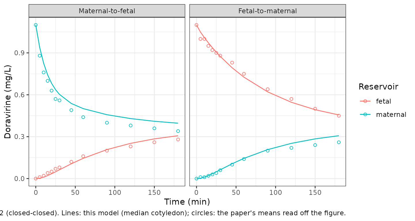
cmp <- sim_long |>
  inner_join(obs_long, by = c("direction", "time", "reservoir")) |>
  filter(time > 0)
cmp_dosed <- cmp |>
  filter((direction == "MTF" & reservoir == "maternal") |
    (direction == "FTM" & reservoir == "fetal"))
stopifnot(
  # Dosed-reservoir decline: a wrong flow, volume or clearance moves the
  # whole curve by tens of percent.
  median(abs(cmp_dosed$sim - cmp_dosed$obs) / cmp_dosed$obs) < 0.12,
  # Whole-figure envelope, both reservoirs, both directions.
  quantile(abs(cmp$sim - cmp$obs), 0.9) < 0.1
)
The transfer shape is reproduced in both directions. The model is for the median cotyledon, whereas the observed curves are means over placentas of 18.9-64.4 g, which is why the dosed reservoir sits slightly off the observed mean late in the fetal-to-maternal experiment.
Closed-open model: configuration sensitivity analysis
The closed-open re-fit lowered CLpdm from 37.2 to 11.0
mL/min and CLpdf from 5.5 to 4.3 mL/min (70% and 22% lower;
Section 3.5), but the 95% CIs overlapped and the impact on the PBPK
predictions was marginal. In the closed-open circuit the dosed
circulation recirculates (200 mL reservoir) while the other is
single-pass, so its collecting reservoir (3 mL) only ever holds a dilute
effluent.
mtf_co <- solve_perfusion(ui_co, ftm = 0L, wt = 33.15)
#> Warning: No sigma parameters in the model
#> ℹ omega/sigma items treated as zero: 'etalcl_pdm', 'etalcl_pdf'
ftm_co <- solve_perfusion(ui_co, ftm = 1L, wt = 33.15)
#> Warning: No sigma parameters in the model
#> ℹ omega/sigma items treated as zero: 'etalcl_pdm', 'etalcl_pdf'

co_end <- tibble::tibble(
  Direction = c("Maternal-to-fetal", "Fetal-to-maternal"),
  `Closed reservoir 180 min` = c(
    mtf_co$Cmaternal[mtf_co$time == 180], ftm_co$Cfetal[ftm_co$time == 180]
  ),
  `Open effluent 180 min` = c(
    mtf_co$Cfetal[mtf_co$time == 180], ftm_co$Cmaternal[ftm_co$time == 180]
  )
)
knitr::kable(co_end, digits = 3)| Direction | Closed reservoir 180 min | Open effluent 180 min |
|---|---|---|
| Maternal-to-fetal | 0.329 | 0.062 |
| Fetal-to-maternal | 0.384 | 0.038 |
stopifnot(
  # The dosed (closed) reservoir declines to a few tenths of the start, and the
  # single-pass open side stays an order of magnitude below it (Online Resource
  # 15 A/B, digitised: closed reservoir ~0.33-0.40, open effluent ~0.03-0.06).
  co_end$`Closed reservoir 180 min` > 0.20,
  co_end$`Closed reservoir 180 min` < 0.50,
  co_end$`Open effluent 180 min` < 0.5 * co_end$`Closed reservoir 180 min`
)
The closed reservoir declines to roughly a third of its starting value while the open-side effluent stays low, matching the Online Resource 15 A/B curves for the two directions.
Between-placenta variability
Both fits carried a between-placenta variability fixed at 100% CV on each transfer clearance. Simulating a cohort of 200 cotyledons in the maternal-to-fetal direction (closed-closed) shows the spread this produces in the fetal reservoir at 180 min.
set.seed(74)
n_cot <- 200
obs_t <- c(0, 60, 120, 180)
ev <- do.call(rbind, lapply(seq_len(n_cot), function(i) {
  d <- rbind(
    data.frame(time = 0, evid = 1L, amt = 192, cmt = "maternal_reservoir", dvid = NA_integer_),
    data.frame(time = obs_t, evid = 0L, amt = 0, cmt = "maternal_reservoir", dvid = 1L)
  )
  d$id <- i
  d
}))
ev$WT_COTYLEDON <- 32.55
ev$PERFUSION_FETAL_TO_MATERNAL <- 0L
sim_pop <- as.data.frame(rxode2::rxSolve(ui_cc, ev, returnType = "data.frame"))
f180 <- sim_pop$Cfetal[sim_pop$time == 180]
summary(f180)
#>    Min. 1st Qu.  Median    Mean 3rd Qu.    Max.
#> 0.04568 0.21540 0.26410 0.24527 0.29044 0.31417
stopifnot(
  # The typical-value fetal reservoir at 180 min (~0.27) sits inside the
  # cohort, and the 100% CV spread is wide.
  median(f180) > 0.15, median(f180) < 0.40,
  stats::IQR(f180) > 0.02
)
NCA
The paper reports no non-compartmental parameters for the ex vivo experiment, so there is no published NCA to compare with. For reference, PKNCA summarises the simulated dosed- and receiving-reservoir exposures over the 180-min maternal-to-fetal closed-closed experiment.
conc <- mtf_fig |>
  pivot_longer(c(Cmaternal, Cfetal), names_to = "reservoir", values_to = "conc") |>
  mutate(id = 1L, reservoir = recode(reservoir, Cmaternal = "maternal", Cfetal = "fetal")) |>
  filter(!is.na(conc))
dose_df <- tibble::tibble(
  id = 1L, time = 0,
  amt = c(220, 0),
  reservoir = c("maternal", "fetal")
)
conc_obj <- PKNCAconc(conc, conc ~ time | reservoir + id)
dose_obj <- PKNCAdose(dose_df, amt ~ time | reservoir + id)
intervals <- data.frame(start = 0, end = 180, cmax = TRUE, tmax = TRUE, auclast = TRUE)
nca <- pk.nca(PKNCAdata(conc_obj, dose_obj, intervals = intervals))
as.data.frame(nca$result) |>
  select(reservoir, PPTESTCD, PPORRES) |>
  pivot_wider(names_from = PPTESTCD, values_from = PPORRES) |>
  dplyr::rename(
    "Reservoir" = reservoir,
    "Cmax (mg/L)" = cmax,
    "Tmax (min)" = tmax,
    "AUC0-180 (min*mg/L)" = auclast
  ) |>
knitr::kable(digits = 2)| Reservoir | AUC0-180 (min*mg/L) | Cmax (mg/L) | Tmax (min) |
|---|---|---|---|
| fetal | 33.00 | 0.31 | 180 |
| maternal | 92.07 | 1.10 | 0 |
Assumptions and deviations
-
Only the ex vivo model is packaged. The whole-body
maternal-fetal pregnancy PBPK model is a Simcyp platform model whose
physiological parameters are database outputs not printed in the paper,
so it is out of scope. The two quantities that bridge the two models,
CLpdmandCLpdf, are in these files. -
Cotyledon weight is a covariate. Table 1 / Online
Resource 17 standardise the placental sub-volumes to a 42 g (44.02 mL)
cotyledon and report
CLpdm/CLpdffor that typical cotyledon (footnote a). The paper scaled volumes and clearances by each placenta’s individual cotyledon weight; this is encoded asWT_COTYLEDON, and the figures above are simulated at the cohort-median weight (32.55 g closed-closed, 33.15 g closed-open). - Diffusion-only structure. The P-glycoprotein transport model (Figure 1B, Equations 3 and 5) was rejected by the authors and is not implemented.
-
Mass balance of the printed equations. Equations
1-7 are written as
dN/dt = (flux terms in N) / Vwith “N denotes amount”. Read literally the reservoir inflow would not balance the outflow; the balances conserve mass only if the right-hand-sideNare concentrations (amount / volume), which is how they are implemented and which reproduces the reported 180-min concentrations. - Observed starting concentration for Figure 3. The Figure 3 overlay doses to the observed starting concentration of about 1.1 mg/L (220 ug in 200 mL) rather than the nominal 0.96 mg/L, so the linear model’s transfer shape overlays the figure directly; the 180-min comparison above uses the nominal 192 ug dose.
- Closed-open values were read off Online Resource 15. The paper reports no end concentrations for the closed-open experiment in the text; the closed-open gate uses the digitised Online Resource 15 A/B curves (reading error about 0.02-0.03 mg/L) and the published direction-specific estimates.